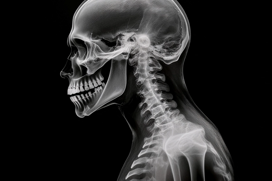
What if there was a way to capture the movement of your organs during everyday activities like breathing or walking? Dynamic Digital Radiography (DDR) is changing the way medical imaging works by allowing healthcare providers to observe organs and muscles in motion providing more detailed insights than traditional X-rays. This article delves into the significance of DDR, its current applications in medical diagnostics and how it is set to transform the future of healthcare. By exploring the benefits and limitations of this innovative technology, we aim to provide readers with a comprehensive understanding of how DDR is reshaping diagnostic practices and improving patient care.
What is DDR?
Dynamic Digital Radiography (DDR) is an advanced imaging technique that allows doctors to see moving images of the body unlike regular X-rays that only capture still images. This technology is particularly useful in examining the diaphragm, the muscle that plays a crucial role in breathing to check for any dysfunctions or abnormalities. Unlike traditional X-rays, which give a snapshot of the body at a single moment, DDR captures a continuous series of images much like a video. This allows doctors to observe how organs and muscles move in real time, providing a clearer picture of how well the diaphragm and other structures are functioning during natural activities such as breathing.
How Does DDR Work?
DDR works by capturing X-ray images at a high speed up to 15 frames per second. These images are then put together to create a moving movie of the body part being examined. For example, when assessing diaphragm function DDR captures how the diaphragm moves during breathing, helping doctors identify any issues with its movement or function. What makes DDR special is that it offers continuous imaging, which allows clinicians to evaluate both structure and function simultaneously. It is particularly helpful for diagnosing diaphragm dysfunctions such as paralysis or weakness, that may not be visible in traditional X-rays.
DDR vs. Traditional X-ray
1. Traditional X-ray:
● Produces a single, still image.
● Primarily focuses on structural issues like fractures or tumors.
● Cannot show how organs or muscles are functioning during movement.
2. Dynamic Digital Radiography:
● Captures real-time motion like a video.
● Allows doctors to see both the structure and function of organs or muscles in action.
● Ideal for diagnosing conditions that affect movement such as diaphragm dysfunction.
Applications of DDR in Medicine
DDR has many uses in diagnosing a range of health conditions particularly in the areas of respiratory health and musculoskeletal issues.
1. Respiratory and Pulmonary Applications
a) Diaphragm Dysfunction:
DDR provides high temporal resolution imaging that allows clinicians to visualize diaphragmatic movement throughout the respiratory cycle. This is particularly beneficial in detecting abnormalities such as unilateral or bilateral diaphragmatic paralysis, paradoxical diaphragm motion and weakened diaphragm activity following surgeries like cardiothoracic procedures or liver transplants. Unlike conventional imaging that captures static views, DDR offers a real-time, non-invasive alternative to fluoroscopy or ultrasound making it easier to assess breathing mechanics and monitor recovery in postoperative patients.
b) Pulmonary Blood Flow Assessment:
DDR is also being utilized to estimate pulmonary perfusion particularly in patients who have undergone lung transplantation or are at risk of pulmonary embolism. By tracking motion-dependent changes in lung opacity during respiration, DDR helps identify perfusion imbalances or vascular complications such as transplant rejection or pulmonary artery obstruction. This dynamic imaging method offers a non-invasive and radiation-efficient alternative to traditional ventilation-perfusion scans enhancing post-transplant monitoring and early detection of issues related to blood flow distribution within the lungs.
c) Chronic Lung Diseases:
In chronic obstructive pulmonary diseases like COPD, emphysema and bronchiectasis, DDR contributes significantly by offering real-time assessment of ventilation and lung movement. It enables visualization of regional airflow patterns and helps detect areas of hyperinflation or impaired lung function. This dynamic insight complements conventional pulmonary function tests (PFTs), allowing for a more comprehensive understanding of disease severity and progression. DDR can also reveal compensatory mechanisms in the lungs supporting personalized treatment plans and ongoing disease management.
d) Airway Collapse and Tracheomalacia:
One of DDR’s emerging uses includes detecting airway collapse during breathing, which is vital in diagnosing conditions such as tracheomalacia or bronchomalacia. The ability to visualize the upper airway dynamically through the respiratory phases makes DDR particularly useful in evaluating these disorders non-invasively, especially in pediatric and elderly populations where traditional methods may be challenging. This technique provides a safe, quick and detailed view of airway dynamics, facilitating timely diagnosis and intervention.
2. Musculoskeletal Applications
a) Joint and Spine Evaluation:
DDR captures motion in joints and the spine, making it a valuable tool for assessing musculoskeletal function. It allows visualization of joint instability, subluxations, spinal misalignments and degenerative changes such as those seen in scoliosis or cervical spine disorders. DDR is especially useful in evaluating injuries like whiplash or disc herniations, where subtle motion abnormalities may not be evident on static imaging. By recording real-time movement DDR supports a more dynamic and functional assessment of musculoskeletal health.
b) Postoperative Monitoring:
In orthopedic recovery, DDR plays a crucial role in monitoring patient progress following surgical procedures such as knee or hip replacements and spinal fusions. It helps in evaluating the alignment and movement of implants and bones as well as assessing rehabilitation outcomes. DDR provides detailed insights into joint range of motion and spinal flexibility which can guide the customization of physiotherapy regimens and support effective recovery planning. The ability to track functionality dynamically adds value to traditional postoperative follow-up methods.
3. Cardiovascular and Neurological Applications
a) Emerging Cardiovascular Uses:
Though still under clinical research, DDR shows promising potential in cardiovascular imaging. It may soon become a useful tool for visualizing vascular and cardiac motion enabling the assessment of conditions like aortic aneurysms, vascular compliance and possibly arrhythmogenic areas within the heart. By capturing pulsatile movements and vessel dynamics, DDR could provide an innovative, non-invasive method for detecting subtle abnormalities that are difficult to assess with standard static radiography.
b) Neurological Assessments:
DDR is also being explored in the evaluation of spinal cord and neurological function. By visualizing spinal biomechanics during load bearing or movement DDR offers insights into dynamic conditions such as syringomyelia, tethered cord syndrome or degenerative spinal diseases. This capability helps in assessing spinal cord dynamics and detecting abnormalities in neural element movement that may not appear in static MRI or CT scans. As such, DDR serves as a supplementary tool in neurological diagnostics particularly when motion-related pathology is suspected.
4. Other and Emerging Applications
a) Swallowing and Esophageal Disorders:
In the field of gastroenterology, DDR is gaining recognition for its ability to evaluate swallowing mechanisms. By capturing real-time motion of the oropharynx and esophagus during ingestion, DDR helps in diagnosing conditions such as dysphagia, aspiration or esophageal motility disorders. It is also valuable in post-stroke patients to assess the safety of swallowing. DDR offers a safer and quicker alternative to traditional barium swallow studies with reduced radiation exposure and the possibility of conducting assessments in outpatient or bedside settings.
b) Gastrointestinal Motility:
Though still an area of active research, DDR is showing potential in the evaluation of gastrointestinal motility. By capturing subtle movements of the intestines, it may help diagnose disorders such as gastroparesis or postoperative ileus. Real-time tracking of bowel motion could become a useful addition to functional gastrointestinal imaging, especially in settings where conventional motility studies are not feasible or accessible.
c) Functional Thoracic Imaging:
DDR also contributes to thoracic imaging by visualizing rib and chest wall movement. This application is especially beneficial in trauma patients such as those with flail chest or thoracic outlet syndrome, where understanding dynamic chest wall mechanics is essential. DDR helps in diagnosing mechanical impairments and guiding treatment strategies by offering a moving picture of the thoracic cage in action.
d) Pediatric Applications:
In pediatrics, DDR is being explored as a safer and more dynamic imaging option compared to fluoroscopy. It is particularly useful in assessing airway malacia, congenital diaphragmatic anomalies or spinal abnormalities like scoliosis. Given its lower radiation exposure and ability to capture motion with minimal patient cooperation, DDR is a valuable addition to pediatric diagnostic tools offering clinicians a detailed yet gentle method for evaluating complex conditions.
Advantages of DDR
● Reduced Radiation Exposure: DDR uses lower doses of radiation than traditional fluoroscopy and CT scans, making it a safer option for patients especially those who need repeated imaging.
● Real-time Functional Imaging: DDR doesn’t just show the structure of organs and muscles, it shows how they move during everyday activities like breathing. This functional insight is crucial for diagnosing conditions that affect movement.
● Improved Diagnosis: Because DDR captures movement, it can provide more detailed and accurate information about conditions like diaphragm dysfunction or joint instability leading to better diagnoses and treatment plans.
● Efficiency: DDR imaging is fast and efficient. It helps doctors make quicker decisions, reducing waiting times for patients and improving the overall healthcare workflow.
Limitations of DDR
● New Technology: DDR is still relatively new, so it may not be available in all healthcare settings. Some hospitals or clinics may not yet have the necessary equipment.
● Not for All Conditions: While DDR is excellent for evaluating movement, it may not be suitable for diagnosing conditions that don’t involve motion like tumors deeply embedded in tissues. In those cases, other imaging techniques like MRI or CT scans may be needed.
● Requires Skilled Operators: For DDR to work properly, the patient must be positioned correctly, and the imaging must be done carefully to avoid errors. The technology still requires skilled technicians and doctors to ensure the best results.
The Future of DDR
As DDR technology continues to evolve, there are exciting possibilities ahead:
● Artificial Intelligence (AI) Integration: AI could help analyze DDR images more quickly and accurately, making it easier to diagnose conditions based on movement patterns.
● Portable DDR Devices: Researchers are working on making DDR equipment more portable which could help doctors diagnose patients in emergency rooms or even in remote locations where traditional imaging equipment is unavailable.
● Wider Clinical Use: DDR’s applications are expected to expand into other areas of medicine such as gastrointestinal, cardiovascular and neurological imaging, where real-time motion could provide valuable diagnostic insights.
Conclusion
Dynamic Digital Radiography (DDR) is revolutionizing diagnostic imaging by bridging the gap between structure and function, offering clinicians the unique ability to observe organs and systems in motion. From detecting diaphragm dysfunction and airway collapse to assessing joint mobility and spinal dynamics, DDR provides real-time, high-resolution imaging that enhances diagnostic accuracy and patient outcomes. Its low radiation exposure, quick image acquisition and growing applications across multiple specialties make it a valuable tool in modern medicine. While limitations such as limited availability and the need for specialized expertise remain, ongoing advancements particularly in artificial intelligence and portable technologies promise to broaden DDR’s clinical impact. As the healthcare landscape continues to embrace more functional, patient-centered diagnostics DDR stands at the forefront of a new era in medical imaging.
References
- Valeria Santibanez, Thomas J. Pisano, Florence X. Doo, Mary Salvatore, Maria Padilla, Norma Braun, Jose Concepcion, Mary M. O’Sullivan. Dynamic Digital Radiography Pulmonary Function Testing: A Machine Learning Lung Study Alternative. CHEST Pulmonary. 2024;2(3):100052. https://www.sciencedirect.com/science/article/pii/S2949789224000187
- Konica Minolta. Dynamic Digital Radiography. Accessed on May 12, 2025. Available from: https://healthcare.konicaminolta.us/radiography/dynamic-digital-radiography
- Hussain ZB, Khawaja SR, Karzon AL, Ahmed AS, Gottschalk MB, Wagner ER. Digital dynamic radiography-a novel diagnostic technique for posterior shoulder instability: a case report. JSES Int. 2023 Mar 23;7(4):523-526. https://pmc.ncbi.nlm.nih.gov/articles/PMC10328772/#sec2
- Cè M, Oliva G, Rabaiotti FL, Macrì L, Zollo S, Aquila A, Cellina M. Portable Dynamic Chest Radiography: Literature Review and Potential Bedside Applications. Med Sci (Basel). 2024 Feb 7;12(1):10. https://pmc.ncbi.nlm.nih.gov/articles/PMC10885043/#sec1-medsci-12-00010
- Hanaoka, J., Yoden, M., Hayashi, K. et al.Dynamic perfusion digital radiography for predicting pulmonary function after lung cancer resection. World J Surg Onc 19, 43 (2021). https://wjso.biomedcentral.com/articles/10.1186/s12957-021-02158-w#citeas
- Komatsu S, Nakamura T, Hoshikawa Y, et al. (April 18, 2025) Dynamic Digital Radiography (DDR) for Pulmonary Blood Flow Assessment After Lung Transplantation: Insights From Two Case Studies. Cureus 17(4): e82485. https://www.cureus.com/articles/345325-dynamic-digital-radiography-ddr-for-pulmonary-blood-flow-assessment-after-lung-transplantation-insights-from-two-case-studies#!/
- Noriaki Wada, Akinori Tsunomori, Takeshi Kubo, Takuya Hino, Akinori Hata, Yoshitake Yamada, Masako Ueyama, Mizuki Nishino, Atsuko Kurosaki, Kousei Ishigami, Shoji Kudoh, Hiroto Hatabu. Assessment of pulmonary function in COPD patients using dynamic digital radiography: A novel approach utilizing lung signal intensity changes during forced breathing. European Journal of Radiology Open. 2024;13:100579. https://www.sciencedirect.com/science/article/pii/S2352047724000340
- Calabrò E, Lisnic T, Cè M, Macrì L, Rabaiotti FL, Cellina M. Dynamic Digital Radiography (DDR) in the Diagnosis of a Diaphragm Dysfunction. Diagnostics. 2025; 15(1):2. https://www.mdpi.com/2075-4418/15/1/2


